A hand holding a cuvette of brightly glowing fluorescent protein in a darkened lab

Alison G. TeboGroup Leader · Janelia Research Campus · HHMI

Making the
invisible
visible.

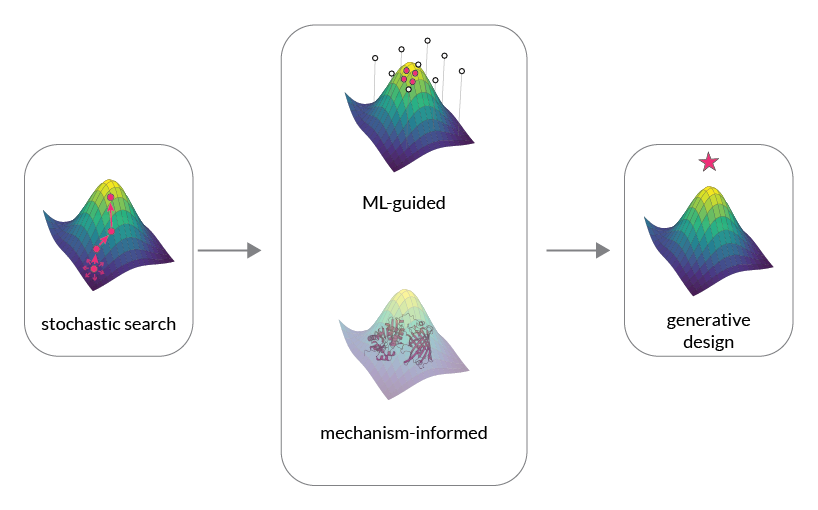
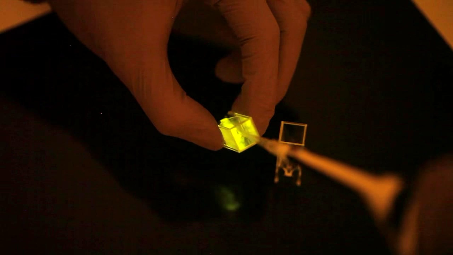
Fluorescent biosensors turn the hidden chemistry of living cells into light — and my lab is building a systematic way to engineer them.

Scroll

The idea

A biosensor is a window. It lets us watch — in real time, inside living systems — when and where the molecules of life interact.

Yet building one is still an art: idiosyncratic, stochastic, one sensor at a time. I want to make it a science.

Why it matters

Seeing life, in color

Calcium spikes. Metabolic flux. Enzyme activity. A biosensor converts these invisible events into fluorescence we can measure — directly, in a living cell.

Give each a different color, and we can watch many at once: a multiplexed, real-time portrait of cellular state.

Green and orange fluorescent protein samples glowing against a black background

Green and red-shifted sensors, side by side — the colors that make multiplexing possible.

The approach

From art to science

An optimized sensor often differs from where it started by only a handful of mutations — yet finding them can take years of trial and error, and the lessons rarely transfer. We change that by combining three things.

High-throughput screening

Explore sequence space at scale and by design — sampling architectures and variants far beyond what hand-picked guesses can reach.

Protein biophysics

Understand the allostery and photophysics that make a sensor work, so improvements come from mechanism rather than luck.

Machine learning

Learn the rules that connect sequence to function — and let models guide which sensors are worth building next.

Diagram of biosensor design moving from stochastic search toward machine-learning-guided, mechanism-informed generative design across a fitness landscape

From stochastic search toward model-guided, mechanism-informed design.

The work so far

Sensors that open
new measurements

Tubes of red fluorescent protein glowing on a red transilluminator

SCaMP

Red calcium indicator

A high-performance red calcium sensor built on mScarlet, engineered through a new topology and directed evolution to deliver the bright, clean red signals that multiplexed imaging has been missing.

Tested in vivo across mouse, zebrafish, fly & worm
Figures showing HYlight2 directed evolution, dose response and improved sensitivity for fructose-1,6-bisphosphate

Population-split directed evolution yields HYlight2.

HYlight2

Glycolysis · fructose-1,6-bisphosphate

A brighter sensor for the sentinel metabolite of glycolysis, evolved by exploring four regions of sequence space in parallel — letting us resolve metabolic dynamics that were previously out of reach.

2.5× the sensitivity of HYlight1

Beneath both sits a quieter result: machine-learning and biophysical models that turn noisy screening data into interpretable, transferable design rules.

What's next

Toward sensors
on demand

01

Scale discovery and optimization

Couple large-scale assays with machine learning, so each round of experiments tells the model what to build next.

02

Ground design in mechanism

Map how sequence sets the allostery between domains and the photophysics of the fluorophore, turning empirical screening into design rules that generalize across sensor classes.

03

Reach new kinds of measurement

Build sensors for the hard targets — protease activity, molecular ratios that reflect pathway flux — using new topologies that current heuristics can't reach.

A biologist who imagines a new measurement should be able to hold a tested sensor within months.

That is the lab we are building.